domingo, 7 de junho de 2015

Interspinous Ligaments May Relate to Chronic Low Back Pain

By Warren Hammer, MS, DC, DABCO
I wonder how many of you bother to palpate spinal interspinous ligaments? I still remember a lecture years ago by James Mennell, MD, whose textbooks on the science and art of joint manipulation are classics, during which he stated that if a ligament is tender upon palpation, there is something wrong with the ligament.
Often through the years I have noticed that some patients with radiating pain extending from their lower back to the buttock and thigh suffer an exacerbation of the pain upon pressure on the C5-C6,C7-C8, L3-L4, L4-L5 interspinous tissue. (Figures 1, 2) Comparing thetender ligament to the one above or below often feels different regarding the density of the tissue. Sometimes, two adjacent ligaments are tender.
Tenderness of the supraspinous ligaments can occur anywhere along the spine. Often, friction massage of the referring ligament reduces or even eliminates the pain. According to Carla Stecco, MD, in her upcoming text, An Atlas of the Human Fascial System, "The superficial fascia adheres to the deep fascia along the spinous processes. In the thoracic region there are many septa never separated by more than 1 mm that insert into the supraspinous ligament. The lumbar region has five fan-shaped thick bundles that originate from the apex of the spinous processes."
cervical interspinal ligaments - Copyright – Stock Photo / Register MarkFig. 1: Pain referral from cervical interspinal ligaments. Patterns of referred pain evoked in normal volunteers by noxious stimulation of interspinous ligaments at the segmental levels indicated. Based on Kellgren.There seems to be evidence that these interspinous ligaments are involved in both acute and chronic spinal pain. M.M. Panjabi theorized that a single trauma or cumulative microtrauma caused subfailure injuries of the ligaments and embedded mechanoreceptors.1"The injured mechanoreceptors generate corrupted transducer signals, which lead to corrupted muscle response patterns produced by the neuromuscular control unit." This can result in muscle incoordination affecting individual muscle force characteristics such as "onset, magnitude, and shut-off."
This situation subjects the individual to abnormal stress and strain in the already aggravated ligaments, mechanoreceptors and muscles exerting increased strain on the facet joints. He states that since spinal ligaments do not have great healing ability, chronic back pain occurs due to eventual inflammation of the neural tissues.1
The corruption of mechanoreceptors is one of the chief hypotheses of fascial manipulation, which attributes the corruption to restricted motion of deep fascia over muscles. The spindle cells, which are located in the fascia, are unable to provide normal feedback to the central nervous system.
This whole concept of ligamentous sensory control changes our attitude about the function of ligaments in general. Ordinarily, we think of ligaments primarily as tissue that maintains the stability of joints. Solomonow, after 25 years of research, makes some (quoted below)2interesting points about ligaments, affirming that ligaments are not passive structures since they exhibit creep, hysteresis and tension relaxation:
  1. "Ligaments are also major sensory organs, capable of monitoring relevant kinesthetic and proprioceptive data.
  2. Excitatory and inhibitory reflex arcs from sensory organs within the ligaments recruit/de-recruit the musculature to participate in maintaining joint stability as needed by the movement type performed.
  3. Long-term exposure of ligaments to static or cyclic loads / movements in a certain dose-duration paradigms consisting of high loads, long loading duration, high number of load repetitions, high frequency or rate of loading and short rest periods develops acute inflammatory responses which require long rest periods to resolve.
  4. Continued exposure of an inflamed ligament to static or cyclic load may result in a chronic inflammation and the associated chronic neuromuscular disorder known as cumulative trauma disorder."
lumbar interspinal ligaments - Copyright – Stock Photo / Register MarkFig. 2: Pain referral from lumbar interspinal ligaments. Patterns of referred pain evoked in normal volunteers by noxious stimulation of interspinous ligaments at the segmental levels indicated. Based on Kellgren.So, applying tension to ligaments stimulates mechanoreceptors and a reflex arc that recruits muscular activity. All ligaments contain Golgi, Pacinian, Ruffini and free-nerve endings. Since it is necessary to rest ligaments, it becomes an argument for lumbar support after an acute injury.
In conclusion, it is important to palpate interspinous ligaments for tenderness and thickening, and attempt to reduce the thickening. It is important to palpate a thickening of restricted fibers before treatment. Some patients are normally hypersensitive and it is essential to rely on palpatory abnormality rather than patient tenderness. Sometimes 2-5 minutes is necessary to palpate a difference.
Often the patient's tenderness will be reduced and the range of motion will improve. The area may or may not require further treatment, but regardless, wait at least 4-5 days before repeating treatment. Again, palpation is more important than tenderness. It also pays to decrease the lumbar lordosis with an abdominal pillow to separate the spinouses.
There are many reasons why mechanical load has a positive effect on soft tissues, and substantial literature supports its use. I find that performing friction massage with my knuckle or using Graston instrumentation works extremely well.
References
  1. Panjabi MM. A hypothesis of chronic back pain: ligament subfailure injuries lead to muscle control dysfunction. Eur Spine J, 2006 May;15(5):668-76.
  2. Solomonow M. Sensory-motor control of ligaments and associated neuromuscular disorders. J Electromyography and Kinesiology, 2006;16:549-567.

Finally, an Accurate Test for a Meniscus Tear?

By Warren Hammer, MS, DC, DABCO
It often appears that when the author of a particular test states high accuracy for the test, other scientists down the line, using MRI or other tests, reach opposite conclusions regarding its validity.
This is certainly true for shoulder labral tests.1 If you've read my previous few articles, you realize this is also probably true for muscle testing. Thus, no matter how logical and accurate any test seems, we must always question it. That is one reason why it pays to use a number of tests to reach any conclusion.
At present, one of the most accurate tests for a knee meniscus tear is palpation of localized tenderness at the joint line, which has an accuracy of 89 percent. An MRI is 98 percent accurate. On the other hand, McMurray is only 58 percent accurate. Other tests, such as Steinman I and II, and the Apley compression and distraction test, are also considered less accurate.
Karachalios, et al., state that based on the evaluation of 213 patients with knee injuries, theirThessaly test is 94 percent accurate in the detection of medial meniscus tears and 96 percent accurate for tears of the lateral meniscus compared to MRI testing.2 After testing, slightly more than 3 percent of the subjects with meniscal tears had an increase in pain after the test that required medication. One patient had a locking of the knee that needed to be treated with manipulation under anesthesia to unlock. None of the controls experienced negative responses to the exam.
Thessaly test - Copyright – Stock Photo / Register MarkThe Thessaly test for meniscus tear evaluation involves external or internal rotation of the body with one foot stabilized straight forward and firmly on the ground.To perform this test, a patient should stand flat-footed with the knee flexed 20 degrees, preferably while holding onto someone or the wall. With one foot stabilized straight forward and the other foot firmly on the ground, the whole body above the knee is either externally or internally rotated (rotation of the femur and the torso in relation to the tibia). See Figures 1 and 2. The test is considered positive when the patient experiences either medial or lateral joint line discomfort or a sense of locking or catching. If the test is positive, it should provoke or reproduce the patient's symptoms.
It is necessary to first perform the test on the normal side. Karachalios, et al., feel that pain occurs due to hoop stress at the intact peripheral rim of the torn meniscus (which is innervated) while loading at 20 degrees of flexion, not because of the application of direct pressure on the torn parts of the meniscus (central portion), which have no nerve endings.
In an article currently in press titled "Validation of the Thessaly test for detecting meniscal tears in anterior cruciate deficient knees," Mirzatolooei, et al., found that the Thessaly test had a low specificity in patients with combined ACL and meniscal injuries, and could not be recommended as a diagnostic test in this association.3 The International Academy of Orthopedic Medicine recommends the Thessaly test as the primary test in the clinical examination to confirm a meniscus tear at the knee. It is a simpler test than the McMurray test. Furthermore, the McMurray test can always be applied in instances of a negative Thessaly test.
The problem is that the average age of subjects in the Mirzatolooei study was 28 years, and no acute knee injuries were included. We also do not know whether this test is accurate if the patient has a history of other knee pathology. Without further research, the test may have limits.
References
  1. Munro W, Healy R. The validity and accuracy of clinical tests used to detect labral pathology of the shoulder: a systematic review. Manual Therapy, 2009;14:119-30.
  2. Karachalios T, Hantes M, Zibis AH, et al. Diagnostic accuracy of a new clinical test for early detection of meniscal tears. J Bone Joint Surg, 2005;87-A(5):955-62.
  3. Mirzatolooei F, Yekta Z, Bayazidchi M, et al. Validation of the Thessaly test for detecting meniscal tears in anterior cruciate deficient knees. Knee, 2009 Sept. 12. [Epub ahead of print]

Overhead Deep Squats: Understanding Movement and Function


By Jeffrey Tucker, DC, DACRB
What are the most common imbalances patients present with? The obvious answer is musculoskeletal imbalances. This article discusses the functional assessment of stability and mobility to movement re-education.
Assessment of the overhead deep squat for stability and mobility imbalances will improve your awareness of the patient's movement dysfunction. Training stability and providing manual mobilization and/or self mobilization are current concepts of movement dysfunction.
A restricted segment can cause a compensation that leads to uncontrolled and increased motion. The uncontrolled segment or region is the most likely site of the source of pathology and symptoms of mechanical origin. Common dysfunctions within the movement system occur when the ankle, hip or thoracic spine needs mobilization, or when the knee, lumbar spine or glenohumeral joint needs stabilization.
There is plenty of evidence to support the link between uncontrolled intersegmental translation or uncontrolled range of motion and the development of musculoskeletal pain and degenerative pathology. Motor control dysfunction within the ankle, knee, hips, lumbar region, thoracic region and shoulder contribute to insidious onset, chronicity and recurrence of pain.
We need to restore ankle dorsiflexion, hip flexion/extension and/or hip adduction/abduction, and thoracic flexion and extension, because there is a frequent relationship between the loss of range of motion at one or more motion segments, and the development of compensatory excessive movement at adjacent segments. Learning to refine mobility and stability will reduce asymmetries and limitations as a means of injury prevention. It is important to establish stabilization prior to strengthening. Evaluate flexibility limitations and asymmetries between the left and right sides of the body. An individual conceivably could overcome a deficit in range of motion in one joint by using more ROM at another joint to achieve the specified goal.
The body is a "kinetic chain" of interconnected parts. I recommend overhead deep squatting as the primary assessment to evaluate whether mobility or stability is required.
The overhead deep squat: The ideal criteria for a well-performed overhead deep squat are:
  1. upper torso parallel with the tibia or toward vertical (back is relatively upright);
  2. femur below horizontal;
  3. knees aligned over feet;
  4. both arms overhead with the dowel aligned over feet;
  5. toes pointed forward; and
  6. knees don't turn in or out.
Hypomobility at any joint in the lower extremity kinetic chain can challenge the motor-control mechanisms of the patient and lead to joint instability. Joint hypomobility can present as dysfunction of intra-articular motion, producing limitations of the accessory movements of roll and glide between the joint surfaces. Limited range of motion also can occur in the myofascial system (extra-articular in nature). These two components are interrelated and often occur together. The abnormal displacement or restrictive barrier to movement changes the normal pattern of movement of the instantaneous axis of rotation (IAR). Movement around an abnormal axis of rotation imposes abnormal compression or impingement on some aspect of the joint tissues and produces altered proprioceptive input to the central nervous system. The motor-control system must adapt to maintain function. These faulty movements increase microtrauma in the tissues around the joint, which, if accumulative, lead to dysfunction and pain.
After an ankle sprain, hypomobility may occur at the subtalar joint, talocrural joint, distal tibiofibular joint, or proximal tibiofibular joint. Limited dorsiflexion after lateral ankle sprain has been attributed to tightness in the gastrocnemius-soleus complex, capsular adhesions developed during immobilization, and subluxations or any combination.
Ankle: The hypomobility of the ankle or tissue tightness can be observed during the overhead deep squat if the heel of the foot rises while descending from a neutral starting position. This is the result of limited soleus muscle motion (e.g., ankle dorsiflexion). Motion can be restored and maintained despite restricted arthrokinematic motion. Restoration of dorsiflexion and normal gait patterns occurs after anterior-to-posterior (manual or self) mobilizations of the talus in the mortise.
If the patient's toes turn outward while descending from the starting position, it means he or she may have weak, tight lateral gastrocnemius, hamstrings, weak inner thighs, and is at risk for Achilles tendonitis.
The progression of rehab to improve the foot dysfunction is to start the patient with ankle self-mobilization. The patient starts out in the double-leg stance. Take a single step forward onto a stool with the right foot. Ask the patient to flex the ankle and knee over the stool as far as they can go. Compare to the left side. The restricted side can be stretched and mobilized while on the stool by repetitively moving the knee over the foot. Altered movement of the subtalar joints and soft tissue tightness can be restored through self repetitive range of motion maneuvers. Next, have the patient perform a wall stretch. With their hands against a wall, feet flat on the ground and one foot at least 18 to 20 inches behind the other, have them bend the front knee. Hold the static stretch for at least 30 seconds. Do this at least two times per leg. The next exercise involves standing on one foot, turned in 45 degrees with the heel hanging off a step. The patient's body weight is on the forefoot. Have them hold onto a wall or rail handle and let their body weight drop down. Instruct the patient to hold this stretch for at least 60 seconds.
Knee: If the knees drift inward while descending from the start position of the overhead deep squat it may mean the patient has weak glutes, tight inner thighs, and is prone to knee and low back problems. The patellofemoral joint may be influenced by the segmental interactions of the lower extremity. Abnormal motions of the tibia and femur in the transverse and frontal planes are believed to have an effect on the patellofemoral joint. The first progression for the knee is to use a foam roll on the adductor and abductor muscles. Firmly press and roll along the tight tissue for several minutes or until you feel a release of tight tissue. Have the patient perform a lunge at a 2 o'clock or 3 o'clock pose with the right leg and a 10 o'clock to 11 o'clock pose with the left leg. The patient should next perform side-lying leg raises. Do not allow the quadratus lumborum muscle to activate early. Raise and lower the top leg, keeping it straight. Isolate the TFL and glute medius. Only perform this on the side that drifts.
Hip: If the patient can keep the feet straight ahead or have only slight external rotation, plus the heels stay flat on the floor while squatting, but they cannot achieve the depth of getting the femurs below the horizontal, they may have tightness where the TFL attach into the glutes. The hip joints may be restricted. The rehab progression is to start with manual mobilization of the hips. Teach the patient how to perform hip range of motion on their own. Part of this solution is simply to do repetitive squats. Over time and many repetitions, the patient will break up the tissue tightness and be able to squat lower and lower.
If you suspect a patient is having a hip extension firing problem during gait, with the hamstrings dominating the movement pattern, rocker sandals can help retrain the gluteus maximus. There are a number of ways to "wake up" the gluts while squatting: for example, weight shift toward the heels, bridges up and down with a therapy band around knees to provide resistance to abduction; side steps with a band around the ankles; or bridges on a gym ball with alternate heel raises. Tight hip flexors will inhibit the gluteus, so these need to be evaluated for length.
For a stronger gluteal contraction, perform the Tucker test, the purpose of which is to help recruit a deeper and stronger contraction of the gluteal group. Test: Place a quarter on the outside of the patient's clothes between the buttocks at the level of the anus, and have the client hold it in place with a strong gluteal contraction. Assess: Can the patient contract the gluteals strong enough and continuously while performing the bridge exercise up and down so the quarter does not drop to the floor? Relate: In order to hold the quarter in place, the patient must concentrate on performing a strong gluteal contraction. This forces the continuous contraction of the gluteus and initiates a co-contraction of the abdominals. Progression: Have the patient perform the overhead deep squat with the quarter held in the buttocks.
Lumbar: If the patient's back bends into flexion while performing the overhead deep squat, it may mean they have tight hip flexors, a weak core and poor posture. This is such an important diagnostic tool. Why is this point so important? The lumbar spine may be more flexible relative to the hips in flexion due to lengthened erector spinae and shortened hamstrings. This can lead to a hamstring strain, but more importantly, the muscles that control excessive lumbar flexion (lumbar erector spinae) have more "give" than the muscles that limit hip flexion (hamstrings). Consequently, during trunk flexion the lumbar spine gives more easily than the hips and excessive flexion occurs in the lumbar spine relative to the amount and time of flexion at the hip joints, resulting in compensatory lumbar flexion and a potential lumbar flexion stability dysfunction. The patient complains of flexion-related symptoms in the lumbar spine. You can see how this will translate to their everyday life. See if you can detect the following possible flexion movement dysfunctions in the low back when the patient forward leans while performing the overhead deep squat:
  1. Shortened back extensor mobilizer muscles (longissimus and iliocostalis): The pelvis shifts more than 4 to 5 inches posteriorly during forward bending and the spine demonstrates limited flexion.
  2. Shortened hamstrings: The hips demonstrate less than 70 degrees of hip flexion during forward bending.
  3. Lengthened gluteus maximus: The hips demonstrate more than 90 degrees of hip flexion during forward bending.
  4. Lengthened back extensor stabilizer muscles (superficial multifidus and spinalis): The spine demonstrates excessive flexion during forward bending.
The progression of rehab is to use the foam roll on the anterior and lateral sides of the hips. Work out as much tissue tightness as you can on the foam roll. To stretch the hip flexors, teach your patient to do a lunge with an arm raised overhead. The precise steps are as follows: Leading with the right foot, the patient performs a lunge while raising the left arm overhead and rotating the upper body to the left. Instruct the patient to hold this pose for 30 seconds and to perform at least two stretches on each side. The most important solution for this movement dysfunction is to control movement at the site of the instability. This concept is a process of sensory-motor re-programming to regain proprioceptive awareness of joint position, muscle activation and movement coordination. This training is beyond the scope of this article. However, you can start by teaching clients to co-contract the mutifidus and transverse abdominus muscles.
Thoracic: During the overhead deep squat, the patient presentation of lack of mobility in the thoracic spine may include the inability to get the dowel directly over the feet. I usually find the arms way out in front of the feet. These patients lack thoracic extension. You will feel restricted motion on palpation of the thoracic spine into extension. The patient may have an obvious forward-drawn posture, anterior head and shoulder carriage (slumping) and/or an increased kyphosis. The rehab solution for this dysfunction is mobilization. The foam roll will allow for self mobilization into extension. The repetition of performing self-mobilization of the thoracic spine into extension, while the patient performs the overhead deep squats, is an exercise in and of itself. Another self-mobilization maneuver involves asking the patient to sit on a chair facing the wall, leaning the forehead on crossed arms against the wall. The patient's knees and toes touch the wall. Taking deep breaths in and out, on the exhalation the patient forces thoracic extension movement, repeating the process about 10 times. I often find the thoracolumbar junction, T6 and above, as the key joints to manipulate to create flexibility.
Shoulder: The gleno-humeral functions. Stability is sacrificed to a large degree to achieve this mobility. During the overhead deep squat you will observe the patient pushing the dowel behind their back instead of over the head. To correct the instability in the shoulder we need to correct the length-tension relationship, improve muscle endurance and coordination of the rotator cuff muscles. These muscles act in a manner to generate a force balance to maintain centering of the joint throughout the range of motion.
Assessment of the overhead deep squat provides analysis of stability and mobility. An exercise program based on the assessment can be implemented to achieve stability and mobility. Stability is only tested reliably under low-load situations. Mobility is based on the ability to pass or fail the ideal criteria of the overhead deep-squat posture. The benefits of having good stability function of both the local and global stabilizer muscles, as well as good joint flexibility, are improved low-threshold motor control and reduced mechanical musculoskeletal pain.
Resources
  1. Bergmark A. Stability of the lumbar spine. A study in mechanical engineering. Acta Orthopaedica Scandinavia 1989;230(60):20-24.
  2. Caterisano A, Moss RF, Pellinger TK, Woodruff K, Lewis VC, Booth W, Khadra T. The effect of back squat depth on the EMG activity of 4 superficial hip and thigh muscles. J Strength Cond Res August 2002;16(30:428-32
  3. Cholewicki and McGill. Mechanical stability in the vivo lumbar spine: implications for injury and chronic low back pain. Clinical Biomechanics 1996;11(1):1-15.
  4. Clark M. "Introduction to Kinetic Chain Dysfunction." Course notes, 2005. Copyright NASM.
  5. Comerford M. "Lumbo-Pelvic Stability." Course notes, 2003. Copyright M. Comerford.
  6. Vermeil A. "Sports & Fitness." Course notes, 2005. Copyright A. Vermeil.

A Nonsurgical Approach for Treating Meniscus Injury

By Warren Hammer, MS, DC, DABCO
Patients occasionally enter the office with a torn meniscus of the knee. In these cases, it is important to determine if they could respond to conservative care. Meniscal tears may be either traumatic or degenerative, and degenerative tears are closely associated with osteoarthritis.
Based on symptomatology, examination and age, one might consider a degenerative meniscal tear from a plain X-ray, but acute tears do not have any specific radiographic findings.1
While magnetic resonance imaging (MRI) is considered the best method for visualizing the knee meniscus, the tear appearing on MRI has no significant basis unless it is based first on the history and physical examination. Between 36-76 percent of asymptomatic patients show tears on MRI and the percentage greatly increases with age. Asymptomatic patients older than age 65 show meniscal tears at a 67 percent rate, and tears are prevalent in 86 percent of patients with symptomatic osteoarthritis.2 Since asymptomatic meniscal tears are common, it is essential that a practitioner be certain that the meniscal tear is the source of the patient's pain.
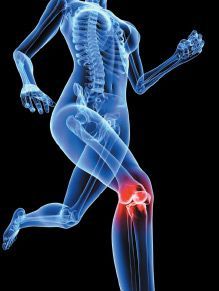
knee pain - Copyright – Stock Photo / Register MarkOne of the most important factors with conservative vs. surgical care is the location of the tear with regard to meniscal vascularity, since the areas of the meniscus with the most vascularity have the greatest ability to heal. The periphery of the menisci is where the blood supply originates (perimeniscal capillary plexus), which derives its supply from the outer medial and lateral geniculate arteries.
Only the peripheral 25-30 percent of the meniscus is vascularized,1 leading to a division of the meniscus with regard to its circulation. The outer third is called the red-red zone, the middle third is called the red-white zone and the remainder of the internal zone (adults) is called the white-white zone. In the red-red zone, bleeding can result in the formation of fibrovascular scar tissue and attract anabolic cells due to cytokines released during the inflammatory phase. The red-white zone has less vascularity and healing potential is therefore less. The white-white zone receives nutrition from synovial fluid by passive diffusion, which can be stimulated by knee joint motion, but since a healing response is not created, the prognosis is poor regarding surgical repair in this zone.
Healing is influenced by the pattern of the tear and the type of vascularity. Longitudinal tears heal better than radial tears. Simple tears heal better than complex tears. Traumatic tears have higher healing rates than degenerative tears, and acute tears heal better than chronic tears.3
Of the many tests used to diagnose a meniscal tear, tenderness at the medial joint line scores highest in terms of accuracy (76-86 percent), the Apley grinding test 46 percent, painful hyperextension 43 percent, Steinmann I sign 42 percent, and McMurray test 35 percent. Most meniscus tests attempt to trap abnormally mobile fragments of the menisci between the femur and the tibia, causing either pain or clicking; but the use of joint-line tenderness in patients with both a meniscus tear and an acute anterior cruciate ligament tear has been shown to be completely unreliable. Joint-line tenderness is most reliable when the tenderness is localized to the posteromedial or posterolateral corner of the knee, since anterior joint-line tenderness is usually present with patellofemoral disease.4
A simple "squat test," whereby the patient raises and lowers themselves from a crouched position, is more indicative of a patellofemoral problem as long as the squat is less than 90 degrees flexion. In 90 degrees knee flexion, 85 percent of the joint load is transmitted through the menisci.4
A particular soft-tissue method I have found beneficial over the years for meniscal tears relates to the treatment of peripheral tears. Free nerve endings of the meniscus are only found on the peripheral one-third, and since palpation of the meniscus at the joint line seems to be the most acceptable test for determining a meniscal lesion, you might assume that the lesion may be more peripheral where the circulation is most prominent.
For the medial joint-line tender area, if it is the right knee, flex the patient's knee 90 degrees and externally rotate the foot and tibia to open up the medial space. Externally rotating the foot also puts pressure on the medial meniscus tear from the medial femoral condyle. Internally rotate the foot and tibia for a lateral tear. Use friction massage with your finger or Graston Technique over the area until you feel a marked decrease in tissue density. (This might take up to 5 minutes.) Do not repeat this treatment for at least five days.
Pain during knee flexion implicates the posterior horns. Pain with extension implicates the anterior horns. Internal rotation tests the lateral meniscus while external rotation tests the medial meniscus.
Manipulation may be performed within the first 24 hours of acute locking (swelling may prevent manipulation) or in chronic locking. Mennell describes a manipulation for a medial-locked torn meniscus as follows.4 He states that in all his years of treating meniscal locking by manipulation, he has never had to treat a lateral lock.
  1. Standing on the supine patient's left side, flex the patient's left knee approximately 110 degrees.
  2. Put your left forearm over the patient's lower tibia and medial malleolus, grasp the calcaneus with the hand and then externally rotate the foot (the entire procedure may also be attempted with the foot internally rotated).
  3. With the right hand, steady the knee with a minor valgus stress. It is important that the tibia remain in a sagittal neutral position (no varus or valgus) during the entire manipulation.
  4. Fully flex the knee, literally kicking the knee into the buttock.
  5. If locking is not immediately reduced, the patient should be referred to an orthopedist.
Estimation of the severity of the lesion, the age of the patient, the degree of knee instability, and the patient's occupation all have significance in determining whether conservative treatment may be attempted. Prolonged loss of knee extension, chronic severe pain, locking, and swelling are definite indicators for possible surgery. An acute injury in young patients (usually in their 20s) should always make one suspect more than just the isolated meniscus lesion. Usually there is pain, swelling, giving way and locking. Often the meniscus tear is associated with an MCL or ACL lesion.
In an athlete, an acute meniscus lesion usually prevents the individual from walking off the field unaided. (This is not the case with an isolated ligamentous injury.) The patient usually complains of a giving way at the time of injury. The meniscus may displace, causing an immediate loss of extension. Patients with medial meniscal tears create a knee locking at 10-30 degrees of flexion, while laterally displaced meniscal tears lock in greater degrees of flexion, especially posterior tears at more than 70 degrees.
Pain at extreme knee extension is affecting the anterior horn, while pain at extreme knee flexion is affecting the posterior horn. The pain in a medial meniscus injury is more often in the posterior medial or medial joint line and is rarely localized anteromedially. Lateral meniscus joint line pain is more often midlateral than posterolateral.4
A degenerative tear of the meniscus in an older patient (40 years or older) is more likely to be an isolated lesion. Older individuals may develop a tear for no apparent reason or feel it as they arise from a chair. If they develop chronic symptoms, they may present with thigh atrophy and weakness associated with pain, effusion, and giving way.
An isolated meniscus lesion will develop mild effusion gradually over a few days, compared with the almost immediate swelling of an anterior cruciate lesion, although swelling can be rapid in a meniscus tear if it occurs in the peripheral vascular zone. If the swelling occurs a day or so later, the tear is probably in the nonvascular central meniscal area. Immediate locking (loss of knee extension) may occur, especially if a bucket-handle tear (longitudinal type) occurs.
Failure to extend the knee may be due to eventual effusion (hamstring spasm and pseudo-locking), so it is important to question the patient to find out whether it was possible to extend the knee fully immediately after the injury. Rarely, a posterior vertical tear of the lateral meniscus will cause a locking in full flexion.4 The majority of the time, knee locking occurs in extension. Of course, rehabilitation, stretching and strengthening should be included with a conservative approach.
References
  1. Mazak TG, Fabricant PD, Wickiewicz TL. Indications for meniscus repair. Clinics in Sports Med, 2012;31(1):1-14.
  2. Greis PE, Bardana DD, Holmstrom MC, et al. Meniscal injury: basic science and evaluation.J Am Acad Orthop Surg 2002;10(3):168-76.
  3. McCarty E, Marx RG. Meniscal tears in the athlete. Operative and nonoperative management. Phys Med and Rehab Clin North Amer, 2000;11(4):867-878.
  4. Hammer W. Functional Soft Tissue Examination and Treatment by Manual Methods, 3rd Edition. Jones & Bartlett: Sudbury, MA, 2007.

Fascial Manipulation

By Marc Heller, DC
Fascial manipulation (FM) is a systematic approach to soft-tissue work evolved over 30 years by Luigi Stecco, an Italian physical therapist.
I'll start out with my thoughts on this treatment approach in the format of a Web review. My rating is 4.5/5 stars; very good.
Pros: It's a unique way of looking at the soft tissues. An amazing map of the body, identifying many significant fascial points that most of us in the soft-tissue world have not previously looked at. A clear-cut end point for the therapeutic soft-tissue session; when the treated points have decreased in tenderness by 50 percent and have markedly softened, you are done. A treatment strategy that, in my limited experience, seems to completely eradicate the fascial densifications. The problem points, once treated, do seem to "hold" and not recur or require multiple treatments.
Again, in my limited experience, this work seems to make a significant clinical difference, helping some of my most chronic patients I was previously unable to help. It is a commitment to an evidence-based approach and to understanding the underlying biological process, with research into fascia and several peer-reviewed papers.
Cons: It is a bit too much of a "technique" for me; the work is taught as a freestanding methodology, without integration of rehab, joint manipulation or other soft-tissue techniques. (I recognize that to bring something new into the world requires singular focus over the long haul; Stecco certainly has that quality.) Additionally, the work can be painful to the patient. I am not saying that one should not do it for that reason, I am just noting a downside.
Further Thoughts
Is this just another variation on soft-tissue / fascial work? Fascial manipulation goes beyond the usual soft-tissue approaches in a couple of ways. One is the extensive dissection studies of unembalmed cadavers that have been done by the Stecco family, well-pictured in his texts. Stecco has studied the fascia in a comprehensive and direct way. FM is not just a new way to push on muscles, but a more comprehensive systemic approach to the influence of soft tissue on pain syndromes in the body. There is a correlation between pain patterns, tender and dense points, and functional testing, with movement and resistive testing to guide your treatment.
The treatment is not necessarily applied to where the patient hurts, but involves a search pattern based on the extensive mapping that Stecco has provided. The emphasis is multifactorial. The doctor is taught to look and test at old trauma sites and see how they may contribute to current fascial problems; and to look above and below the site of pain. The doctor is also reminded to look at the antagonist muscles and fascia.
Stecco has also extensively reviewed the existing literature on fascia and connective tissue. His large body of work includes several books, including two in English, and multiple articles in peer-reviewed journals. He has many new hypotheses about what we are doing to soft tissue, and how it works. He is not just a another technique guru, but one of those rare practitioners who has both a scientific mind and is a profound clinical innovator.
I have always been attracted to and appreciated systems that recognize pain is a liar. My interest in applied kinesiology, visceral manipulation and other systems has been in search of the Holy Grail: What is underneath this pain? I think and hope that Stecco has really advanced our understanding of what causes pain and what we can do to help effectively treat chronic musculoskeletal pain.
Stecco says search the fascia in a more comprehensive way. Once you have found the significant points, completely erase them. I have always done my soft tissue quickly. I assumed that once I initiated a change, once I began to feel the soft tissues melt or change, the body could complete the process. In FM, first you find a dense and tender point or small area that correlates with the functional testing and the pain pattern. You then treat it with a combination of 80 percent compression and 20 percent oscillation until the tenderness has diminished at least 50 percent, and the density of the knot is gone. This process, which is painful to the patient, takes between two and five minutes.
This work, like most deep-tissue techniques, is both painful and pro-inflammatory. This requires explanation to the patient. It is OK if they are sore or even hurt like heck the next day. The Steccos have looked at the literature and have fascinating evidence-based theories about this component. One, there is a cycle of about 48 hours of inflammation that centers around the hyaluronic acid system, found in the ground substance. I don't fully understand the science, but what I do understand is that when the soft tissues are deeply manipulated, you have initiated an inflammatory process that contributes to healing of the tissues; a process that peaks at about 24 hours and is mostly gone within 48 hours. I have observed this phenomenon while doing Graston Technique; it is great to have further biological explanation of what is happening at the cellular level.
The mapping of the fascia and its correlations with specific pain patterns and specific functional tests is amazing. This is an amazing body of work, clearly the work of a lifetime. This is not just a tweak, but a new way to look at the musculoskeletal system.
An Example: Knee Pain
An example will help. Anterior knee pain, patellar pain, is certainly a common problem in athletes and aging people. We have many tools for this already, including rehab, ART, Graston Technique, and others. Stecco noted a specific point, in the anterior quadriceps at the junction of the rectus femoris and the vastus lateralis, that seems to correlate with this pain pattern. If the patient has difficulty with a deep lunge or squat, and this point is tender and dense, you treat it with the deep fascial manipulation. The patient will frequently have an immediate improvement in their function. Of course, there are other fascial points that one would search and potentially treat, but this one is key. I have almost never focused on this area previously for knee pain.
This is not just anecdotal, Pedrelli, Stecco, et al., wrote a peer-reviewed article on this particular issue ["Treating Patellar Tendinopathy With Fascial Manipulation." J Bodyw Mov Ther, Jan 2009;13(1):73-80]. One of the fascinating things about this article, this study, is that it represents a dumbing down, an oversimplification of the Stecco model. In the complete FM model, one would treat more than one point, and assess anterior knee pain via various functional tests. The study just treated the one usual main point, in the upper quadriceps, and got great results anyway.
A Couple of Cases
I'll extensively quote a colleague of mine, Phillip Snell, DC, another recent student of FM, to comment further on the knee and on fascial manipulation. Dr. Snell both tells his own clinical case story and looks at why this may work.
"The interplay and the inherent limitations of manual therapy vs. functional rehab exercise was apparent to me recently at a Fascial Manipulation (Stecco) seminar. I had chronic anterior knee pain with an orthopedic diagnosis of 'osteoarthritis.' I looked for unconventional solutions after ART, 'quad sets' and manipulation were not productive. About this time, Graston Technique came on the scene, and I was able to get 80 percent relief in ADLs using it on myself.
"In recent years, functional assessment brought a diagnosis of patellofemoral syndrome, and rehab working on the gluteals got another 10 percent function. My litmus test for whether I felt my knee was whole (as I approach 50 with a history of ACL surgery) was whether I could do a deep single-leg squat, a pistol. Single-leg variations of squats, deadlifts and hip thrusts still resulted in intermittent pain in the knee. (In behavioral medicine, intermittent reinforcement is one of the most effective ways to inculcate behavior.) Thus, I had a 'hard deck' of about 70 degree depth on my single-leg squat for several years. That is, until the Stecco class.
"While attending a Fascial Manipulation seminar, a couple of points in the hip adductors and TFL of my affected knee were positive on assessment. After applying FM techniques for 15 minutes, I dropped into not one, but 4-5 consecutive assisted single-leg pistols! Mind you, I had been doing gluteal rehab, working on mobility in the hip flexors and adductors, and using my kit bag of myofascial release techniques on those areas for over a year."
[Author's note: Phillip was working at the table right behind me. I turned around and was amazed to see him comfortably in this extreme single-leg squat position; I wish I had a picture.)]
"Such quick results are almost certainly neurological in nature. FM work appears to eliminate the neurological inhibitors. In this case, I think a possible cause of the limited motion was possible shearing of free nerve endings in densified fascia. This may have produced local pain and/or decentration of joints; so the brain would not allow that full movement pattern."
I will share one more case history. This is a 20-year-old female, who was in an auto accident two years prior. She had chronic, 8/10 thoracic pain, with no changes on MRI. She was referred to me as a last resort by her orthopedist. I did all of my magic, all of my thinking outside of the box, for eight weeks. I cleared her anterior cervical spine, I addressed the thoracic and rib-cage restrictions. I did anterior visceral manipulation. I found lumbosacral issues and corrected those.
She came in, just after I had taken this FM seminar, for her ninth visit, and was quite frustrated. She still was in severe pain, still flared up whenever she was active at all. I suggested we try a couple of sessions of FM. Despite my inexperience in this technique, I somehow found the right points. Two treatments and her pain levels came down to a 2-3/10, and she seemed like a different person. I thank you, Luigi Stecco, and so does this patient.
What is the take-home from this article for you, the practicing DC? One, if you can, study fascial manipulation. If not, remember to look above and below the pain site, look at the antagonist muscles and fascia, not just the site of pain. With patients in which the same fascial restrictions or tight tender knots recur, consider treating these points longer and deeper, until you completely erase the hot spots. Don't forget to warn the patient that they will be sore.